Bilayer mucoadhesive intrauterine delivery platform for sustained platelet-derived extracellular vesicle release to promote endometrial regeneration and improve implantation outcomes
DOI:
https://doi.org/10.69857/joapr.v14i4.2180Keywords:
Extracellular vesicles, Endometrial receptivity, Mucoadhesive biomaterials, Intrauterine delivery, Regenerative therapy, ImplantationAbstract
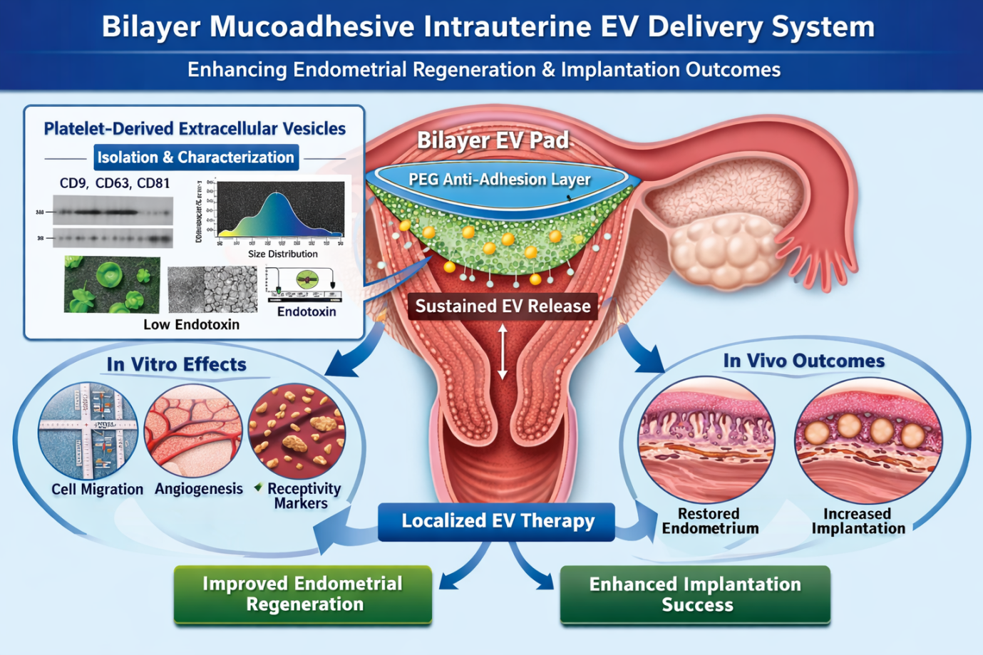
Background: Endometrial dysfunction remains a major cause of implantation failure and infertility. Extracellular vesicles (EVs) derived from platelet-rich plasma have shown regenerative potential; however, their clinical application is limited by rapid clearance and poor retention within the uterine cavity. Methodology: A bilayer mucoadhesive intrauterine delivery platform was developed for sustained release of platelet-derived extracellular vesicles. The bilayer system consisted of a mucoadhesive EV-loaded layer and a protective anti-adhesion layer. Physicochemical characterization, EV release kinetics, and mucoadhesion properties were evaluated. In vitro wound healing, cell proliferation, and gene expression studies were performed. In vivo efficacy was assessed through histological evaluation of endometrial thickness and implantation outcomes. Results and Discussion: The bilayer platform demonstrated sustained extracellular vesicle release for up to 120 h with minimal initial burst release. Enhanced cell migration and proliferation, along with upregulation of endometrial receptivity markers including LIF, HOXA10, Integrin αV/β3, and IGFBP1, were observed. In vivo studies revealed significant improvement in endometrial thickness and implantation sites in the EV-Pad group compared to controls. The sustained release of EVs improved endometrial regeneration and enhanced implantation potential. The bilayer design demonstrated improved local retention characteristics and enhanced regenerative responses compared with free EV administration under experimental conditions. Conclusion: The developed bilayer mucoadhesive intrauterine delivery system provides a promising strategy for sustained EV delivery and improved endometrial regeneration, supporting its potential as a preclinical platform for sustained intrauterine regenerative delivery.
Downloads

References
Zhang X, Wei H. Role of decidual natural killer cells in human pregnancy and related pregnancy complications. Front. Immunol., 12, 728291 (2021) https://doi.org/10.3389/fimmu.2021.728291
Jia Y, Liu Y, Li Y, Guan X, Xu J, Yan Z, et al. Persistent Wnt signaling affects IVF embryo implantation and offspring metabolism. Sci. Bull., 70, 2297–2311 (2025) https://doi.org/10.1016/j.scib.2025.05.003
Lessey BA, Young SL. What exactly is endometrial receptivity? Fertil. Steril., 111, 611–617 (2019) https://doi.org/10.1016/j.fertnstert.2019.02.009
Altmäe S, Esteban FJ, Stavreus-Evers A, Simón C, Giudice LC, Horcajadas JA, et al. Endometrial transcriptomics. Hum. Reprod. Update, 26, 707–722 (2020) https://doi.org/10.1093/humupd/dmaa020
Vaidakis D, Papapanou M, Siristatidis CS. Autologous platelet-rich plasma for assisted reproduction. Cochrane Database Syst. Rev., 4, CD013875 (2024) https://doi.org/10.1002/14651858.CD013875.pub2
Ouyang Y, Mouillet JF, Coyne CB, Sadovsky Y. Placenta extracellular vesicles in pregnancy. Am. J. Reprod. Immunol., 83, e13204 (2020) https://doi.org/10.1111/aji.13204
Obuchi W, Zargani-Piccardi A, Leandro K, Rufino-Ramos D, Di Lanni E, Frederick DM, et al. Engineering of CD63 enables selective extracellular vesicle cargo loading and enhanced payload delivery. J. Extracell. Vesicles, 14, e70094 (2025) https://doi.org/10.1002/jev2.70094
Langer R. Drug delivery and targeting. Nature, 392, 5–10 (1998) https://doi.org/10.1038/32381
Park K. Controlled drug delivery systems. J. Control. Release, 190, 3–8 (2014) https://doi.org/10.1016/j.jconrel.2014.03.054
Kalluri R, LeBleu VS. The biology and biomedical applications of exosomes. Science, 367, eaau6977 (2020) https://doi.org/10.1126/science.aau6977
Doyle LM, Wang MZ. Overview of extracellular vesicles. Cell, 181, 1043–1051 (2020) https://doi.org/10.1016/j.cell.2020.04.002
Maas SLN, Breakefield XO, Weaver AM. Extracellular vesicles: Intercellular delivery vehicles. Trends Cell Biol., 30, 172–188 (2020) https://doi.org/10.1016/j.tcb.2019.11.003
Gurung S, Perocheau D, Touramanidou L, Baruteau J. The exosome journey: From biogenesis to uptake and intracellular signalling. Cell Commun. Signal., 19, 47 (2021) https://doi.org/10.1186/s12964-021-00730-9
Pegtel DM, Gould SJ. Exosomes. Annu. Rev. Biochem., 88, 487–514 (2019) https://doi.org/10.1146/annurev-biochem-013118-111902
Witwer KW, Goberdhan DCI, O'Driscoll L, Théry C, Welsh JA, Blenkiron C, et al. Minimal information for studies of extracellular vesicles 2021 (MISEV2021): From basic to advanced approaches. J. Extracell. Vesicles, 10, e12070 (2021) https://doi.org/10.1002/jev2.12070
Bian S, Zhang L, Duan L, Wang X, Min Y, Yu H, et al. Extracellular vesicles derived from stem cells in tissue repair and regeneration. Signal Transduct. Target. Ther., 5, 242 (2020) https://doi.org/10.1038/s41392-020-00353-x
Gupta D, Zickler AM, El Andaloussi S. Dosing extracellular vesicles. Adv. Drug Deliv. Rev., 178, 113961 (2021) https://doi.org/10.1016/j.addr.2021.113961
Herrmann IK, Wood MJA, Fuhrmann G. Extracellular vesicles as a next-generation drug delivery platform. Nat. Nanotechnol., 16, 748–759 (2021) https://doi.org/10.1038/s41565-021-00931-2
Ding JY, Chen MJ, Wu LF, Shu GF, Fang SJ, Li ZY, et al. Mesenchymal stem cell-derived extracellular vesicles in skin wound healing: Roles, opportunities and challenges. Mil. Med. Res., 10, 36 (2023) https://doi.org/10.1186/s40779-023-00472-w
Burnouf T, Goubran HA, Chou ML. Platelet vesicles: Biological functions and applications. Blood Rev., 42, 100709 (2020) https://doi.org/10.1016/j.blre.2020.100709
Yao C, Wang C. Platelet-derived extracellular vesicles for drug delivery. Biomater. Sci., 11, 5758–5768 (2023) https://doi.org/10.1039/D3BM00893B
Wang Y, Tang Z, Teng X. New advances in the treatment of thin endometrium. Front. Endocrinol. (Lausanne)., 15, 1269382 (2024) https://doi.org/10.3389/fendo.2024.1269382
Murphy DE, de Jong OG. Extracellular vesicle therapeutics: Progress and challenges. Nat. Rev. Drug Discov., 22, 415–437 (2023) https://doi.org/10.1038/s41573-023-00641-y
Shelke PV, Rachh PR, Mankar S, Amin S, Jain D. Optimization and evaluation of nebivolol hydrochloride loaded transferosomes using Box-Behnken experimental design. J. Appl. Pharm. Res., 12(4), 124–138 (2024) https://doi.org/10.69857/joapr.v12i4.590
Jafernik K, Ładniak A, Blicharska E, Czarnek K, Ekiert H, Wiącek AE, et al. Chitosan-based nanoparticles as effective drug delivery systems: A review. Molecules, 28, 1963 (2023) https://doi.org/10.3390/molecules28041963
Dash M, Chiellini F, Ottenbrite RM, Chiellini E. Chitosan—A versatile semi-synthetic polymer in biomedical applications. Prog. Polym. Sci., 36, 981–1014 (2011) https://doi.org/10.1016/j.progpolymsci.2011.02.001
Highley CB, Prestwich GD. Hyaluronic acid hydrogels for biomedical applications. Curr. Opin. Biotechnol., 40, 35–40 (2016) https://doi.org/10.1016/j.copbio.2016.02.008
Khutoryanskiy VV. Advances in mucoadhesion and mucoadhesive polymers. Macromol. Biosci., 21, e2000398 (2021) https://doi.org/10.1002/mabi.202000398
Andrews GP, Laverty TP, Jones DS. Mucoadhesive polymeric platforms for controlled drug delivery. Eur. J. Pharm. Biopharm., 71, 505–518 (2009) https://doi.org/10.1016/j.ejpb.2008.09.028
Knop K, Hoogenboom R, Fischer D, Schubert US. Poly(ethylene glycol) in drug delivery: Pros and cons as well as potential alternatives. Angew. Chem. Int. Ed., 49, 6288–6308 (2010) https://doi.org/10.1002/anie.200902672
Jokerst JV, Lobovkina T, Zare RN, Gambhir SS. Nanoparticle PEGylation for imaging and therapy. Nanomedicine, 6, 715–728 (2011) https://doi.org/10.2217/nnm.10.19
Peppas NA, Bures P, Leobandung W, Ichikawa H. Hydrogels in pharmaceutical formulations. Eur. J. Pharm. Biopharm., 50, 27–46 (2000) https://doi.org/10.1016/S0939-6411(00)00090-4
Hoffman AS. Hydrogels for biomedical applications. Adv. Drug Deliv. Rev., 64, 18–23 (2012) https://doi.org/10.1016/j.addr.2012.09.010
Hade MD, Suire CN, Suo Z. Mesenchymal stem cell-derived exosomes: Applications in regenerative medicine. Cells, 10, 1959 (2021) https://doi.org/10.3390/cells10081959
Livshits MA, Khomyakova E, Evtushenko EG, Lazarev VN, Kulemin NA, Semina SE, et al. Isolation of exosomes by differential centrifugation and ultrafiltration. Sci. Rep., 5, 17319 (2015) https://doi.org/10.1038/srep17319
Böing AN, van der Pol E, Grootemaat AE, Coumans FAW, Sturk A, Nieuwland R. Single-step isolation of extracellular vesicles by size-exclusion chromatography. J. Extracell. Vesicles, 3, 23430 (2014) https://doi.org/10.3402/jev.v3.23430
Dragovic RA, Gardiner C, Brooks AS, Tannetta DS, Ferguson DJP, Hole P, et al. Sizing and phenotyping of cellular vesicles using nanoparticle tracking analysis. Nanomedicine, 7, 780–788 (2011) https://doi.org/10.1016/j.nano.2011.04.003
Vyas KS, Kaufman J, Munavalli GS, Robertson K, Behfar A, Wyles SP. Exosomes: The latest in regenerative aesthetics. Regen. Med., 18, 181–194 (2023) https://doi.org/10.2217/rme-2022-0134
Powles TB, van der Heijden MS, Loriot Y, Bedke J, Valderrama BP, Iyer G, et al. Enfortumab vedotin plus pembrolizumab in untreated locally advanced or metastatic urothelial carcinoma: 2.5-year median follow-up of the phase III EV-302/KEYNOTE-A39 trial. Ann. Oncol., 36, 1212–1219 (2025) https://doi.org/10.1016/j.annonc.2025.05.536
Williams KL. Endotoxin detection methods. In: Methods Mol. Biol., 1601, 3–12 (2017) https://doi.org/10.1007/978-1-4939-6960-9_1
Boateng JS, Matthews KH, Stevens HNE, Eccleston GM. Wound healing dressings and drug delivery systems: A review. J. Pharm. Sci., 97, 2892–2923 (2008) https://doi.org/10.1002/jps.21210
Burdick JA, Prestwich GD. Hyaluronic acid hydrogels for biomedical applications. Adv. Mater., 23, H41–H56 (2011) https://doi.org/10.1002/adma.201003963
Smart JD. The basics and underlying mechanisms of mucoadhesion. Adv. Drug Deliv. Rev., 57, 1556–1568 (2005) https://doi.org/10.1016/j.addr.2005.07.001
Cheng L, Hill AF. Therapeutically harnessing extracellular vesicles. Nat. Rev. Drug Discov., 21, 379–399 (2022) https://doi.org/10.1038/s41573-022-00410-w
Galsky MD, Hoimes CJ, Necchi A, Shore N, Witjes JA, Steinberg G, et al. Perioperative pembrolizumab therapy in muscle-invasive bladder cancer: Phase III KEYNOTE-866 and KEYNOTE-905/EV-303. Future Oncol., 17, 3137–3150 (2021) https://doi.org/10.2217/fon-2021-0273
van der Heijden MS, Powles T, Gupta S, Loriot Y, Galsky MD, Valderrama BP, et al. Exploratory subgroup analyses of EV-302: A phase III global study to evaluate enfortumab vedotin in combination with pembrolizumab versus chemotherapy in previously untreated locally advanced or metastatic urothelial carcinoma. ESMO Open, 10, 105544 (2025) https://doi.org/10.1016/j.esmoop.2025.105544
Liang CC, Park AY, Guan JL. In vitro scratch assay: A convenient and inexpensive method for analysis of cell migration in vitro. Nat. Protoc., 2, 329–333 (2007) https://doi.org/10.1038/nprot.2007.30
Arnaoutova I, Kleinman HK. In vitro angiogenesis: Endothelial cell tube formation assay. Nat. Protoc., 5, 628–635 (2010) https://doi.org/10.1038/nprot.2010.6
Goligher EC, Heath A, Harhay MO, Marshall JC, Thabane L, Rubenfeld GD, et al. Bayesian statistics for clinical research. Lancet, 404, 1067–1076 (2024) https://doi.org/10.1016/S0140-6736(24)01295-9
Ruxton GD. The unequal variance t-test is an underused alternative to Student's t-test and the Mann–Whitney U test. Behav. Ecol., 19, 690–693 (2008) https://doi.org/10.1093/beheco/arn020
Pang R, Arbelaiz J, Pillow JW. Learning neural dynamics through instructive signals. bioRxiv (2025) https://doi.org/10.1101/2025.08.30.673300
Calderon Martinez E, Ghattas Hasbun PE, Salolin Vargas VP, García-González OY, Fermin Madera MD, Rueda Capistrán DE, et al. A comprehensive guide to conduct a systematic review and meta-analysis in medical research. Medicine (Baltimore)., 104, e41868 (2025) https://doi.org/10.1097/MD.0000000000041868
Published
How to Cite
Issue
Section
Copyright (c) 2026 Rajaganapathy Kaliyaperumal, Srinivasan R, Kalaivanan Seeni, Vignesh Sekar

This work is licensed under a Creative Commons Attribution-NonCommercial 4.0 International License.